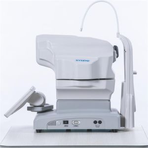
Overview
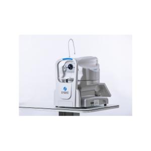
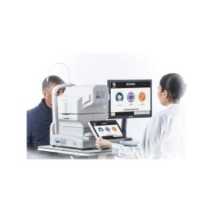
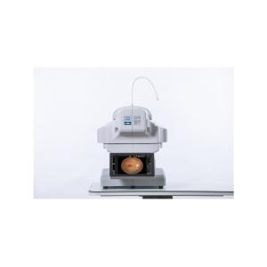
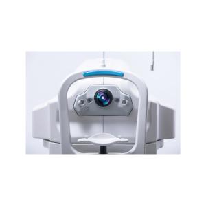
SYSEYE RetiCam 3100 is a non-mydriatic digital fundus camera that captures clear retinal images with small pupils ≥3.3 mm and a 50° field of view. Auto/manual focus and XYZ auto-alignment make operation simple and fast for routine screening of DR, AMD and glaucoma. Standard configuration ships in 5 working days; MOQ = 1.
4 Key Benefits
-
Small-pupil imaging: ≥3.3 mm without dilation
-
50° field of view for routine screening and documentation
-
Easy to use: auto/manual focus with XYZ auto-alignment
-
Fast delivery & low MOQ: ships in 5 days; MOQ = 1
Quick Enquiry (reply within 12 hours)
Please share: (1) Your organization (Clinic/Hospital/Distributor) (2) Country/Region (3) Quantity & target delivery date (4) WhatsApp or Email. We’ll send distributor/volume pricing and arrange a 15-min live demo.
Standard Accessories
Main unit (RetiCam 3100), chin-rest paper pad, power cable, objective lens cover, dust cover, wireless keyboard & mouse, hood, extension plug, operation manual.
Key Specifications
-
Field of view: 50° (typical)
-
Minimum pupil: ≥3.3 mm (small-pupil mode)
-
Resolution (lp/mm): center ≥60; edge ≥25 (reference)
-
Magnification: 1.3×
-
Working distance: 35 mm
-
Focus: auto/manual; refractive compensation ±25 D
-
Alignment: XYZ auto-alignment
-
Illumination: IR LED for observation/alignment; xenon flash for capture
-
Fixation: internal LED; external fixation supported
-
Movement range: stage 90 mm (L/R), 35 mm (F/B); main unit vertical 30 mm; chin-rest 60 mm
-
Power: AC 100–240 V, 50/60 Hz
-
Dimensions/Weight: 380×550×475 mm / approx. 26.5 kg
Compliance & Connectivity
-
Image export: DICOM-compatible formats supported for system integration/archiving.
-
Quality system: ISO 13485. (If FDA 510(k) is available, list the K-number here and upload the certificate; otherwise do not claim FDA.)
Applications
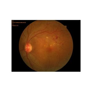
Screening and follow-up for DR/AMD/glaucoma; routine check-ups and referral documentation; education and case management.

| Field of view | 50° |
| Field of view tolerance | ±7% |
| Resolution |
|
| Center of view | ≥60 lp/mm |
| Field of view center (r/2) | ≥40 lp/mm |
| At the edge of the field of view (r) | ≥25 lp/mm |
| Magnification | 1.3 times |
| Required pupil diameter | 4.0 mm or more (3.3 mm or more when using the small pupil shooting function) |
| Working distance | 35 mm |
| Focus adjustment range | ±25D |
| Shooting light | Auto: with shooting mode |
| Manual: can be set manually |
| light source |
|
| Illumination light | Infrared led |
| Shooting light | Xenon lights |
| reflected light |
|
| Scattered light |
|
| camera | Digital camera |
| Fixing light | Internal fixing light (LED) |
| External fixation light |
| Moving range |
|
| Stage | 90 mm left and right, 35 mm front and rear |
| Main unit moves vertically | 30 mm |
| Chin-rest tray movement range | 60 mm |
| Rated power supply | AC 100V~240V,50/60Hz |
| size | 380 mm(L)x 550 mm(W)x 475 mm(H) |
| weight | About 26.5kg |
| When in maximum light intensity/hole bar state |
| Spectral wavelength | 305 nm~1100 nm |